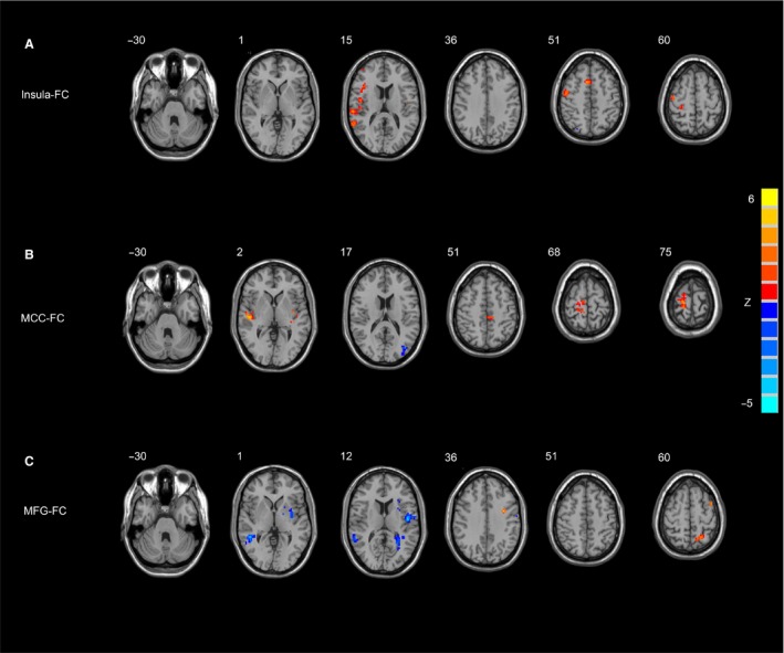
Figure 2.

Significant differences in the FC of each ROI between PD‐F and PD‐NF group. (A) the left insula FC between. PD‐F and PD‐NF group; (B) the right MCC FC between PD‐F and PD‐NF group; (C) the right MFG FC between PD‐F and PD‐NF group. Results are displayed at p < 0.05 corrected by AlphaSim. PD‐F, Parkinson's disease patients with fatigue; PD‐NF, Parkinson's disease patients without fatigue; FC, functional connectivity; MCC, midcingulate cortex; MFG, middle frontal gyrus.
