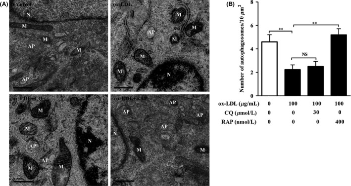
Figure 3.

ox‐LDL resulted in the ultrastructure damage to HT‐22 cells. TEM was used to observe ultrastructure alteration in ox‐LDL‐treated HT‐22 cells. (A) HT‐22 cells were treated with ox‐LDL (100 μg/mL) in the presence of chloroquine (30 μM) or rapamycin (400 nM) for 24 hours. Mitochondria (M), nucleus (N), and autophagosome (AP) were indicated. (B) Average number of APs was quantified (n=10 cells/group). Values are the mean ± SEM. NS, no significant difference. **P<.01. Scale bar, 500 nm
