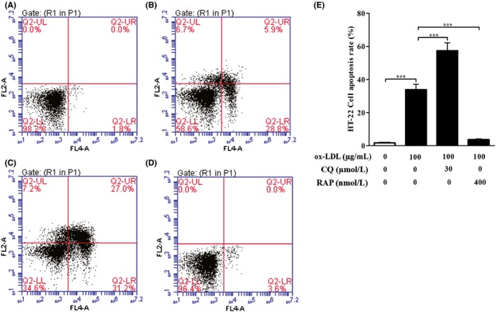
Figure 5.

Effect of autophagy on HT‐22 cell apoptosis induced by ox‐LDL. Flow cytometry was used to measure the effect of autophagy on HT‐22 cell apoptosis induced by ox‐LDL. (A–D) HT‐22 cells were treated with ox‐LDL (100 μg/mL) in the presence of chloroquine (30 μM) or rapamycin (400 nM) for 24 hours. Apoptosis of HT‐22 cells was assessed by flow cytometry as described in 2 section. (A) Control (untreated) group; (B) ox‐LDL group; (C) ox‐LDL + CQ group; (D) ox‐LDL + RAP group. (E) HT‐22 cell apoptotic rates were quantified as described in 2 section. All the data were shown as mean ± SEM of three independent experiments. *** P<.001
