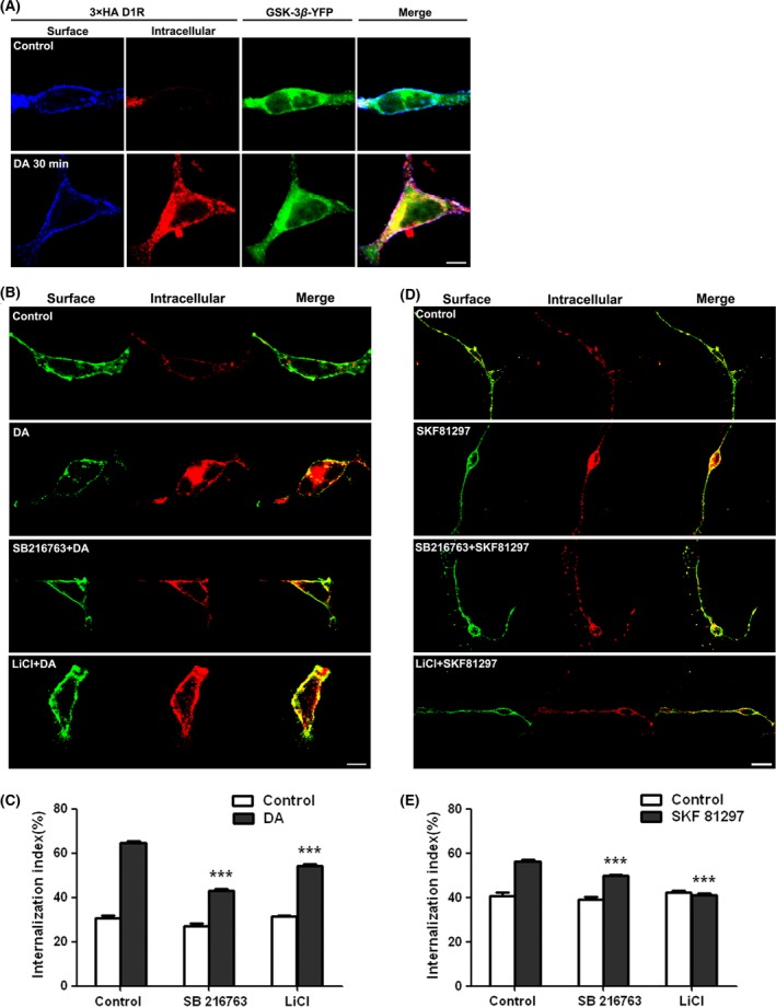
Figure 4.

Inhibition of D1R internalization in HEK‐293‐D1R cells and in primary‐cultured striatal neurons by GSK‐3β inhibitors. (A) Co‐internalization of GSK‐3β with D1R. Stable HEK‐293‐D1R (HA‐tagged) cells were transiently transfected with GSK‐3β‐YFP. Thirty‐six hours after transfection, surface receptors were live‐conjugated with an anti‐HA antibody and were allowed for endocytosis (37°C, 30 min) in the absence or presence of DA (10 μM). (B) A representative image for DA‐stimulated D1R endocytosis in the presence or absence of GSK‐3β inhibitors 5 mM LiCI or 5 μM SB216763 in HEK‐293 cells stably expressing D1R. (D) The quantification for SKF81297‐stimulated D1R endocytosis in the presence or absence of GSK‐3β inhibitors in primary‐cultured striatal neurons transfected with HA‐D1R. (C) and (E) quantitation of the intracellular accumulation assays, measured as the ratio of internalized/total fluorescence (internalization index). Histograms show mean ± SEM (n = 30–35 for each condition). (***P < 0.001, LiCl/SB216763 vs. control).
