
Figure 6.
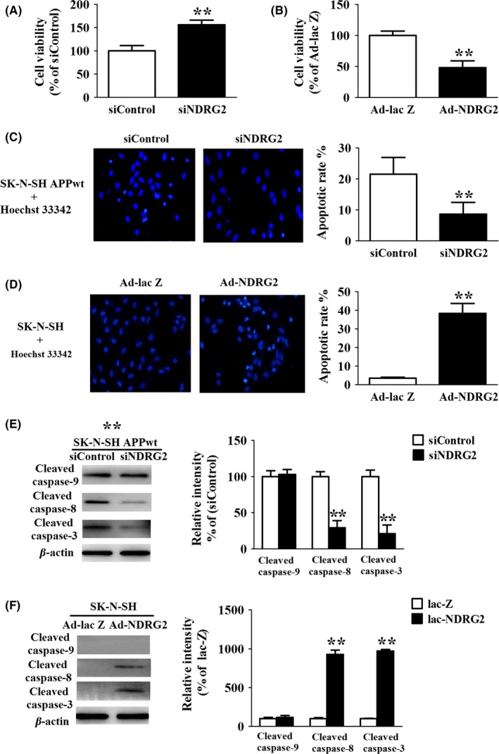
NDRG2 contributes to apoptosis via the extrinsic apoptotic pathways (A) MTT assay of SK‐N‐SH APPwt cells after cells were transfected with siControl and siNDRG2 for 48 h. (B) MTT assay of SK‐N‐SH cells after cells were transfected with Ad‐NDRG2 and Ad‐lacZ for 48 h. (C) Hoechst staining of SK‐N‐SH APPwt cells after treated with siControl and siNDRG2 for 48 h. (D) Hoechst staining of SK‐N‐SH APP cells after treated with Ad‐NDRG2 and Ad‐lacZ for 48 h. (E) Representative Western blots and quantitative analysis of cleaved caspase‐9, cleaved caspase‐8, cleaved caspase‐3 after SK‐N‐SH APPwt cells were transfected with siControl and siNDRG2. (F) Representative Western blots and quantitative analysis of cleaved caspase‐9, cleaved caspase‐8, cleaved caspase‐3 after SK‐N‐SH cells were transfected with Ad‐NDRG2 and Ad‐lacZ for 48 h. Quantified results were normalized to β‐actin expression. For all the results above, a representative experiment of three performed is shown. Values were expressed as percentages compared to the control group (set to 100%) and represented as group mean±SEM. n=4~5 per group. *P<0.05, **P<0.01 vs control group