
Figure 2.
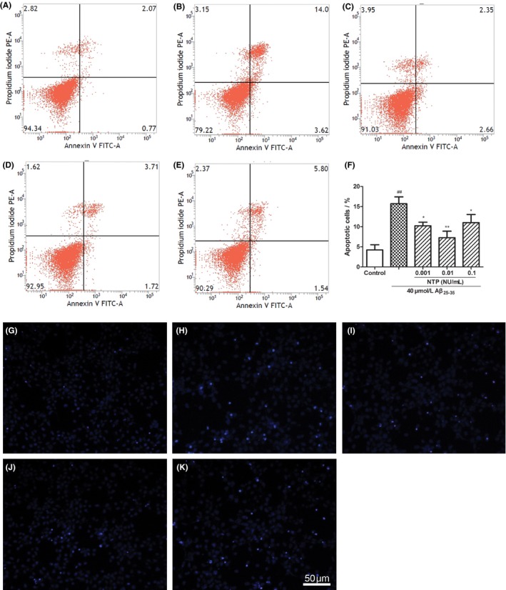
Neurotropin® (NTP) alleviated Aβ25‐35‐induced apoptosis. (A) Control group. (B) HT22 cells were exposed to 40 μmol/L Aβ25‐35 for 24 h. (C‐E) HT22 cells were coincubated with 40 μmol/L Aβ25‐35 for 24 h after pretreatment with various concentrations of NTP (0.001, 0.01, 0.1 UN/mL) for 16 h. Cell apoptosis was assessed by flow cytometry. (F) Statistical results of cell apoptotic rates. (G‐K) HT22 cells were stained with Hoechst 33342 and propidium iodide (PI) and observed under a fluoroscent microscope after treatment. As demonstrated in the pictures, Aβ25‐35 induced apoptosis was characterized by condensed, intensely fluorescent nuclei. Neurotropin® markedly reduced the number of apoptotic cells. Values were represented as relative percentage to the control group and shown as mean±SE (n=6). ## P<.01 versus control, *P<.05 and **P<.01 versus Aβ25‐35 group