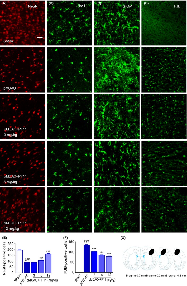
Figure 2.

PF11 rescues pMCAO‐induced neural cell death and glial cell activation. Immunofluorescence images showing that PF11 significantly reversed the pMCAO‐induced neuron death (A), microglial activation (B), and astrocyte activation (C) in the ipsilateral cortex, as detected by laser confocal microscopy using antibodies against NeuN (red), Iba‐1 (green), and GFAP (green), respectively. Fluoro‐Jade B (FJB) staining was used to identify degenerating neuronal cells (D, green). Scale bar=20 μm. (E, F) Statistical analysis of surviving neurons (NeuN‐positive) and degenerating neurons (FJB‐positive) was conducted using Image‐Pro Plus 6.0. The values are presented as means±SEM (n=18). Statistical comparisons were carried out with ANOVA followed by Tukey's test. ###P<.001 vs sham group, ***P<.001 vs pMCAO group. pMCAO denotes rats subjected to pMCAO for 24 h; pMCAO+PF11 (3, 6, 12 mg/kg, iv) denotes rats that were injected with PF11 at 0.5 h after the onset of pMCAO. Rats were perfused with 4% paraformaldehyde, and 20 μm brain cortex sections were processed for immunofluorescence examination. (G) The black ovals indicate the regions selected for immunofluorescence detection
