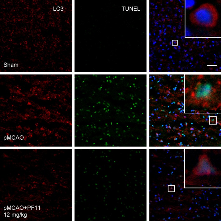
Figure 3.

PF11 attenuates pMCAO‐induced accumulations of autophasosomes and apoptosis. Representative immunofluorescence staining of double‐labeled brain cortex sections showing LC3‐positive cells (red) and TUNEL‐positive cells (green) in sham‐operated rats and pMCAO rats treated with saline or PF11 (12 mg/kg) at 0.5 h after the onset of pMCAO. Nuclei are labeled with DAPI (blue). High‐magnification images of the boxed areas are shown in the inserts. Scale bar=20 μm
