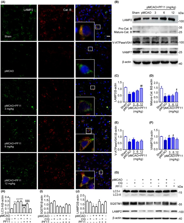
Figure 5.

PF11 attenuates pMCAO‐induced lysosome dysfunction and promotes the fusion of lysosomes with autophagosomes. (A) Immunofluorescence studies showing the colocalization of LAMP2‐positive lysosomes (green) with the lysosomal enzyme CatB (red) in the cortex of sham‐operated rats and of pMCAO rats 24 h after treatment with saline or PF11 (3, 6, 12 mg/kg) at 0.5 h after the onset of pMCAO. Nuclei are stained with DAPI (blue). High‐magnification images of the boxed areas are shown in the inserts. Scale bar=10 μm. (B) Immunoblots showing the levels of LAMP2, mature‐CatB, V‐ATPase V0A1, and VAMP7 in brain cortex extracts from sham‐operated rats and of pMCAO rats 24 h after treatment with saline or PF11 (3, 6, 12 mg/kg) at 0.5 h after the onset of pMCAO. β‐actin was used as a loading control. (C‐F) Densitometric analysis of the immunoblots. Statistical comparisons were carried out with ANOVA followed by Tukey's test. Data are presented as means±SEM. **P<.01, *P<.05 vs sham group; ##P<.01, #P<.05 vs pMCAO group; n=4. (G‐J) Effects of CQ treatment on proteins in the autophagic‐lysosomal pathway. Brain cortex samples were obtained from sham‐operated rats with or without CQ, pMCAO‐operated rats with or without CQ, pMCAO+PF11‐treated rats with or without CQ, and rats treated with PF11 alone. Western blot analysis showing the levels of LC3, SQSTM1, and LAMP2 (G) and their ratio normalized to the β‐actin loading control (H‐J). CQ was administered 5 h before the rats were killed. Statistical comparisons were carried out with ANOVA followed by Tukey's test. Data are presented as means±SEM. ***P<.001, **P<.01, *P<.05 vs sham group; ###P<.001 vs pMCAO group; $$$P<.001 vs pMCAO+PF11 group; n=4
