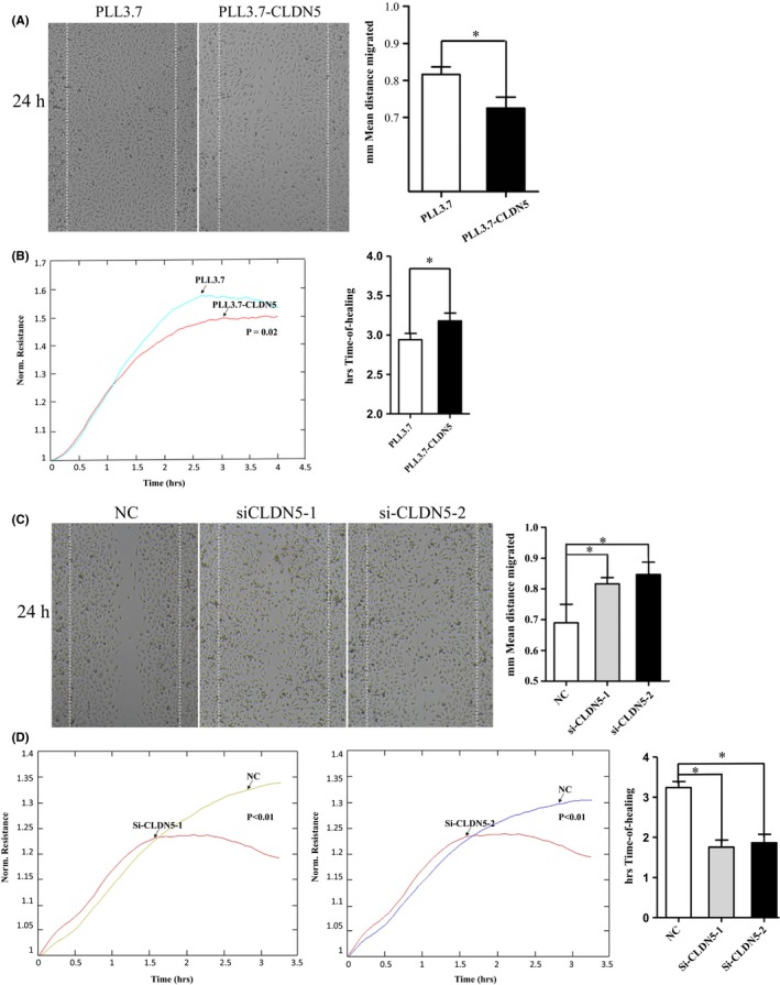
Figure 3.

CLDN5 inhibited hCMEC/D3 cell migration. PLL3.7‐CLDN5 and PLL3.7 cells were subjected to wound healing assays. (A‐B) Larger gap was found in PLL3.7‐CLDN5 cells comparing to the control cells. The impaired motility of PLL3.7‐CLDN5 cells was also verified using electric cell impedance sensing (ECIS), more time was needed for PLL3.7‐CLDN5 cells to reach confluence after wounding created on the panel in ECIS assays. (C‐D) SiCLDN5 cells were more vigorous compared with the control ones. The siCLDN5 cells showed an increased migration rate comparing with the control group in that fewer time was needed for healing. Original magnification, 10×. Values are mean ± SEM from three independent experiments. Statistical significance is indicated
