
Figure 2.
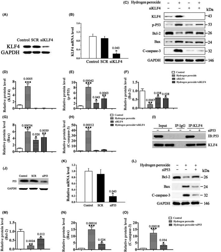
H2O2 induces p53‐dependent RGC apoptosis through upregulation of KLF4. (A, B) The mRNA (B) and protein (A) expression of KLF4 in SCR or siKLF4‐transfected RGCs by RT‐PCR and Western blot analysis, respectively. Untransfected cells were included as control. Data were normalized to GAPDH. n = 3, *P < 0.05 vs. control. (C–H) The protein levels of KLF4, p‐p53, and apoptosis‐related proteins (Bcl‐2, Bax, and cleaved caspase‐3 (C‐caspase‐3)) by Western blot analysis. RGCs transfected with siKLF4 or SCR were treated with 500 μM H2O2 or vehicle for 12 h. Data were normalized to GAPDH. n = 3, *P < 0.05, **P < 0.01, ***P < 0.001 vs. control. (I) Coimmunoprecipitation analysis of the interaction between KLF4 and p53 in RGCs. IgG was used as a control. (J, K) The mRNA (K) and protein (J) expression of p53 in SCR or sip53‐transfected RGCs by RT‐PCR and Western blot analysis, respectively. Untransfected cells were included as control. Data were normalized to GAPDH. n = 3, *P < 0.05 vs. control. (L–O) The protein levels of apoptosis‐related proteins Bcl‐2, Bax, and C‐caspase‐3 by Western blot analysis. RGCs transfected with sip53 or SCR were treated with 500 μM H2O2 or vehicle for 12 h. Data were normalized to GAPDH. n = 3, *P < 0.05, **P < 0.01, ***P < 0.001 vs. control.