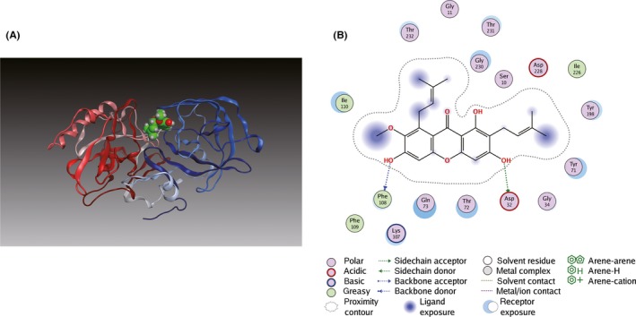
Figure 5.

Binding mode of α‐M to BACE1. (A) α‐M was docked into crystal structure of BACE1 (PDB ID: 1FKN). The N‐terminal lobe and the C‐terminal lobe of BACE1 are blue and red, respectively. (B) Key hydrogen bonding interactions between α‐M and BACE1 at the catalytic residue Asp32 and at Phe108 of N‐terminal lobe are indicated with green and blue dashed lines
