
Figure 1.
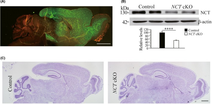
Conditional inactivation of NCT in the forebrain of NCT cKO mice. A, The expression pattern of Cre recombinase. Sagittal brain section was obtained from CaMKIIα‐Cre;mTmG mouse. Green fluorescence (by GFP) is clearly seen in the cortex, the hippocampus, the olfactory bulb and the striatum but not cerebellum. Scale bar = 500 μm. B, Western blotting on NCT using cortical samples of 3‐month NCT cKO mice. There was significant difference on protein levels of NCT between NCT cKO mice and age‐matched littermate controls (control = 100% ± 9.4%, cKO = 50.2% ± 3.0%; n = 3 per group; ****, P < .001). C, Nissl staining. Comparable brain morphology was detected between control and NCT cKO mice. Scale bar = 500 μm