
Figure 6.
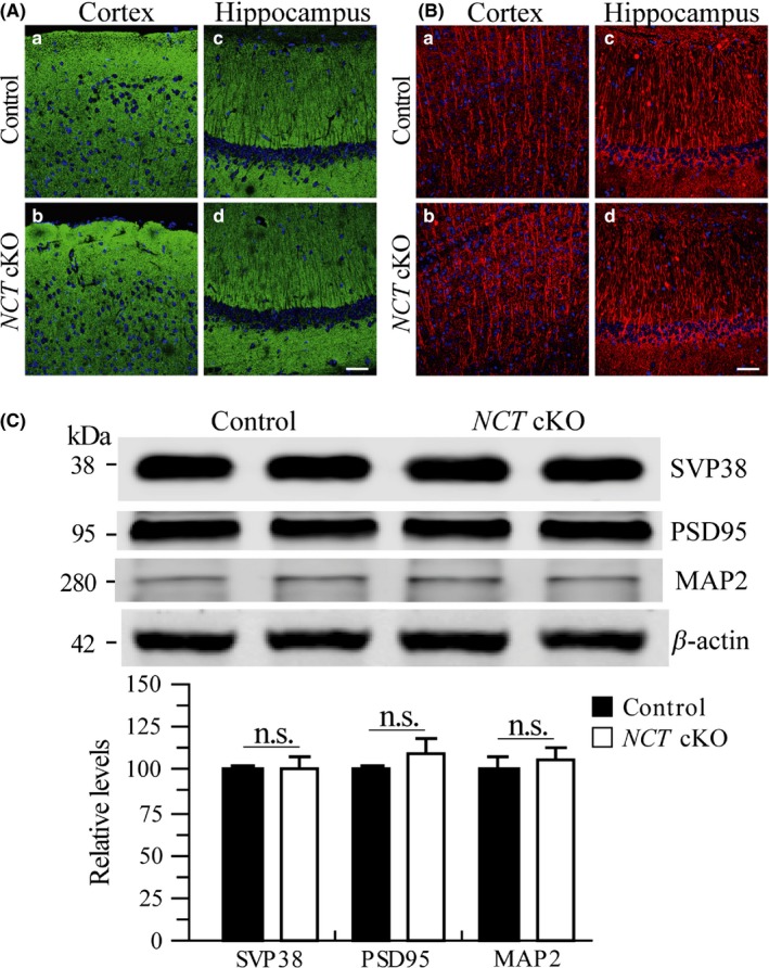
Neither synaptic loss nor dendritic degeneration in NCT cKO mice at 3 mos. A, Immunostaining of SVP38 in NCT cKO mice at 3 months. DAPI+ cells were in Blue, and SVP38 + cells were in Green. There was no detectable difference on SVP38 immunoreactivity in the cortex (a,b) and the hippocampus (c,d) of control and NCT cKO mice. Scale bar = 50 μm. B, Immunostaining of MAP2 in NCT cKO mice at 3 mos. DAPI+ cells were in Blue, and MAP2 + dendrites were in Red. There was no qualitative difference on MAP2 immunoreactivity in the cortex (a,b) and the hippocampus (c,d) of control and NCT cKO mice. Scale bar = 50 μm. C, Western analyses on SVP38, PSD95, and MAP2. There were no significant differences on relative protein levels between control and NCT cKO mice at 3 mos (SVP38: control = 100 ± 3.1%, cKO=101.4 ± 5.5%; PSD95: control = 100 ± 2.7%, cKO = 110.1 ± 7.3%; MAP2: control = 100 ± 8.4%, cKO = 106.8 ± 5.4%; n = 4/group; n.s.: not significant, P > .2)