
Figure 1.
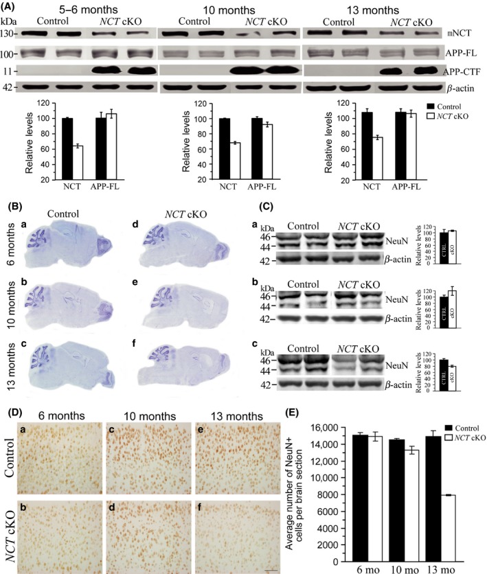
Age‐dependent loss of mature neurons in forebrain‐specific NCT cKO mice. (A) Biochemical analyses on NCT, APP‐FL, and APP‐CTF in NCT cKO mice across ages (n = 3–4/group). Western blotting confirmed significantly decreased levels of NCT in three cKO groups (5–6 months: control = 100 ± 1.2%, cKO = 64.3 ± 2.6%, P = 0.00009, two‐tailed Student t‐test; 10 months: control = 100 ± 0.6%, cKO = 68.0 ± 1.7%, P = 0.003; 13 months: control = 100 ± 4.4%, cKO = 69.9 ± 4.2%, P = 0.007). Western blotting on APP‐FL showed unchanged levels in NCT cKO groups at 5–6 (P = 0.56), 10 (P = 0.11), or 13 (P = 0.81) months of age. Western results for APP‐CTF indicated massive accumulation in NCT cKO mice aged at 5–6, 10, or 13 months. (B) Nissl staining for NCT cKO mice using brain sections aged at 6, 10, and 13 months. The cortex morphology and the cortex size were normal in NCT cKOs at 6 months (a,d). The cortex became smaller in NCT cKOs at 10 months (b,e). The cortex size was further reduced, and the lateral ventricle became bigger in NCT cKOs at 13 months (c,f). (C) Western analysis on NeuN. There were no significant differences on protein levels of NeuN in NCT cKO brains at 5–6 (a: P = 0.55) or 10 (b: P = 0.10) months of age. NeuN levels were significantly decreased in NCT cKO mice at 13 months, as compared to age‐matched littermate controls (c: P = 0.01). (D) NeuN immunostaining. There was no difference on immuno‐reactivity of NeuN in NCT cKO brains at 6 (a–b) and 10 (c–d) months. Immuno‐reactivity of NeuN in NCT cKO brains was decreased at 13 months (e–f). Scale bar=100 μm. (E) Quantification data on the average number of cortical NeuN+ cells per brain section. There was no significant difference between NCT cKOs and controls at 6 (P = 0.82) or 10 (P = 0.13) months. However, the average number of cortical NeuN+ cells was significantly decreased in NCT cKO mice at 13 months (P = 0.009).