
Figure 2.
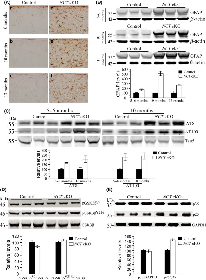
Astroglial activation and tau hyperphosphorylation in NCT cKO mice. (A) Immunostaining of GFAP. GFAP+ cells were intensively seen in the cortex of NCT cKO mice at 6 (a,d), 10 (b,e), and 13 (c,f) months of age, but were hardly detected in age‐matched controls. Scale bar = 40 μm. (B) Western analysis on GFAP. Protein levels of GFAP were significantly increased in NCT cKO mice at 5–6 (P = 0.008, two‐tailed Student t‐test), 10 (P = 0.00002), and 13 months (P = 0.00001), as compared to age‐matched littermate controls. (C) Western blotting on p‐tau using antibodies of AT8 and AT100. Levels of p‐tau were significantly increased in the cortex of NCT cKO mice at 5–6 (For AT8: P = 0.004; For AT100: P = 0.04) and 10 months (For AT8: P = 0.031; For AT100: P = 0.02). (D) Western analyses on GSK3β and p‐GSK3β. Relative levels of p‐GSK3β S9 (P = 0.7) and p‐GSK3β Y216 (P = 0.4) were not decreased in the cortex of NCT cKO mice. (E) Western analyses on p25 and p35. Levels of p25 but not p35 were increased in NCT cKO mice (P = 0.0006).