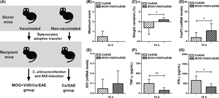
Figure 6.

Splenocytes from vaccinated mice transfer protection against EAE development. Experimental design (A); vaccination procedure included eight VitD doses administered every other day and two MOG doses injected at days ‐16 and ‐8. One day after last VitD dose, splenocytes were adoptively transferred (1.5 × 10⁶ cells/mouse) into C57BL/6‐naïve mice. Recipient mice were infected with C. albicans 3 days before EAE induction. Disease development was followed during the early clinical EAE phase to assess maximum clinical score (B) and percentage of weight variation (C). Expression of FoxP3 (D) and IDO (E) were analyzed in lumbar spinal cord. The TNF‐α (F) and IFN‐γ (G) levels were measured in CNS cell culture stimulated with MOG. The results are expressed as mean ± SD (6 mice/group), and one experiment is shown. *P < 0.05 and **P < 0.01 indicate difference between groups.
