Figure 4.

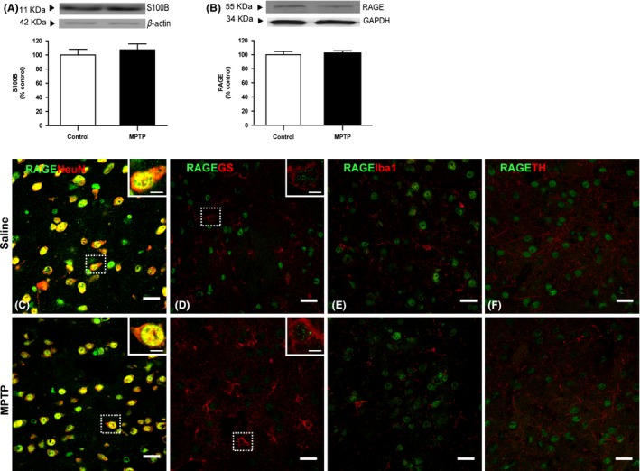
Effect of chronic MPTP treatment on S100B/RAGE axis. Physiological levels of both (A) S100B and (B) mRAGE proteins were observed in striatal total homogenates of MPTP‐treated animals. Double immunofluorescent staining for RAGE (green) and neuronal marker NeuN, astrocytic GS, microglial Iba1, or TH‐positive fibers (red) in striata of saline‐ and MPTP‐treated mice was performed. mRAGE was found to be highly expressed in striatal neurons, showing a punctate distribution mostly localized in nuclei and perikarya of NeuN‐positive cells (colabeling is in yellow) in both saline‐ and MPTP‐treated animals (C). RAGE immunostaining was also found in soma of GS‐positive cells in both saline‐ and MPTP‐treated mice (D), regardless of the astrocytic reactive status. RAGE does not seem to be localized in Iba1‐positive cells (E) nor in TH‐positive fibers from both saline‐ and MPTP‐injured striata (F). S100B and RAGE protein levels were assessed by Western blot, normalized with β‐actin/GAPDH levels, and expressed as % of control. Results are expressed as mean ± SEM of 4 animals per group. Confocal images are representative of four animals per group. Scale bar 20 μm and 5 μm for zoomed images.
