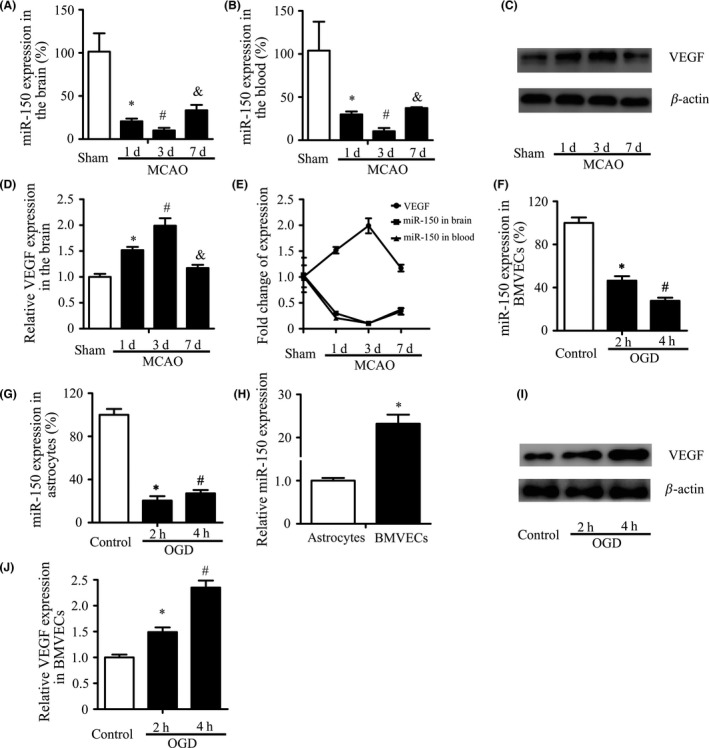
Figure 1.

MiR‐150 is Downregulated after Ischemia. (A–B) MiR‐150 expression decreased significantly following ischemia in brain and blood. Sprague Dawley rats were randomly assigned to four groups (sham, 1, 3, 7 days after MCAO), n = 5. U6 and miR‐16 were used as the internal control, respectively, and data of miR‐150 expression in the brain were normalized by those of the contralateral side. Data were related to the sham group and showed as mean ± standard deviation (SD). *P < 0.05 vs. the sham control, # P < 0.05 vs. 1 day, & P < 0.05 vs. 3 day. (C–E) The expression of VEGF protein in the ischemic border zone (IBZ) was increased following ischemia and negatively related to miR‐150. β‐Actin was used as the internal control. Data were related to the sham group and showed as mean ± standard deviation (SD). *P < 0.05 vs. the sham control, # P < 0.05 vs. 1 day, & P < 0.05 vs. 3 day. (F–G) The expression of miR‐150 in BMVECs and astrocytes decreased significantly. Brain microvascular endothelial cells (BMVECs) and astrocytes were treated with oxygen–glucose deprivation (OGD) for 2, 4 h. U6 was used as the internal control. *P < 0.05 vs. the control, # P < 0.05 vs.2 h. (H) MiR‐150 expression in BMVECs was much higher than in astrocytes. U6 was used as the internal control. *P < 0.05. (I–J) The expression of VEGF protein in BMVECs was increased significantly after OGD. β‐Actin was used as the internal control. *P < 0.05 vs. the control, # P < 0.05 vs. 2 h.
