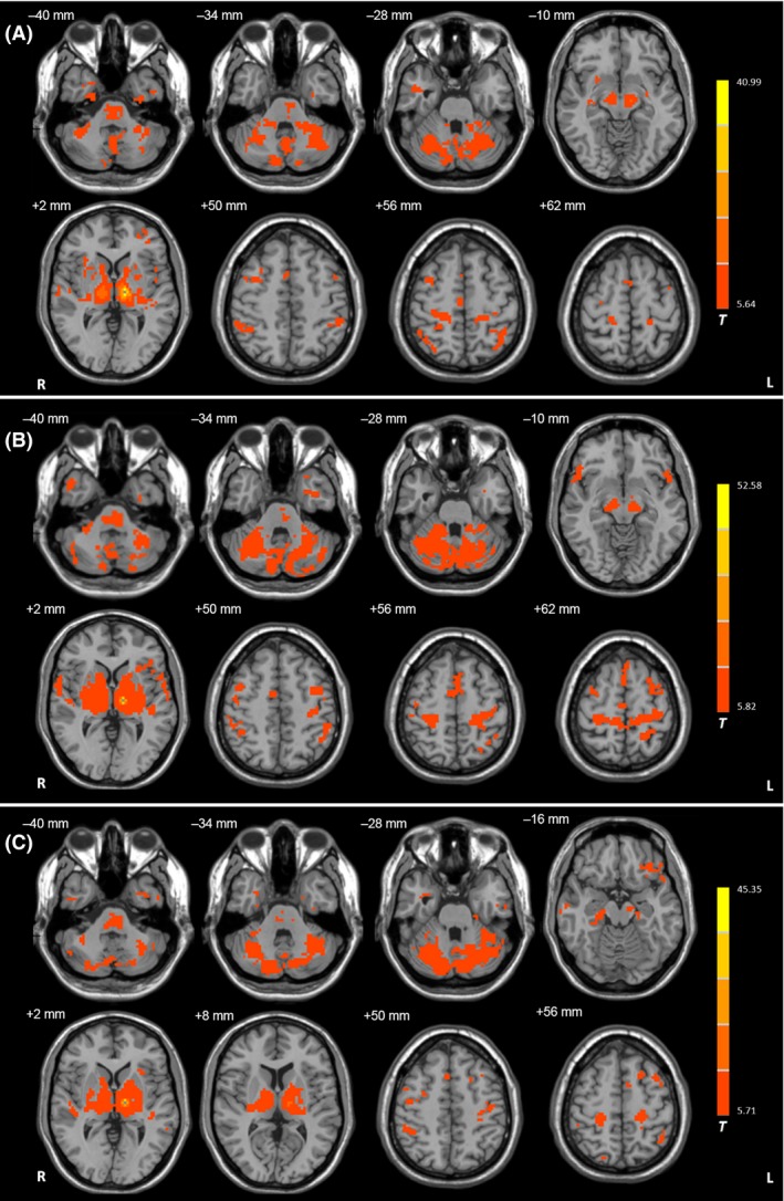
Figure 1.

Functional connectivity in the left Vim nucleus. Brain regions showing significant connectivity with the left Vim nucleus in the resting state in (A) healthy control subjects, (B) tremor‐dominant (TD), and (C) akinetic‐/rigid‐dominant (ARD) patients (one‐sample t‐test, P < 1 × 10⁻⁵, FDR corrected). T value bar is shown on the right. Abbreviations: L, left; R, right.
