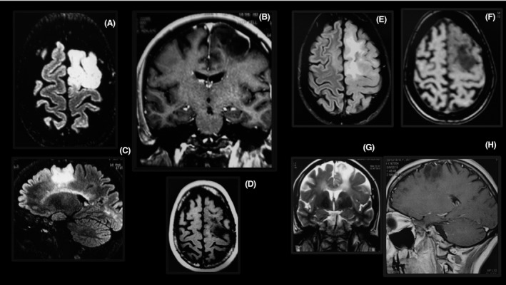
Figure 2.

Tumefactive lesion in the brain of a patient using fingolimod. The lesion is large (A–D), enhanced by gadolinium forming a ring (B), and restricted to the white matter (A–C). Two months after fingolimod withdrawal, the lesions were markedly reduced (E–H).
