Figure 4.

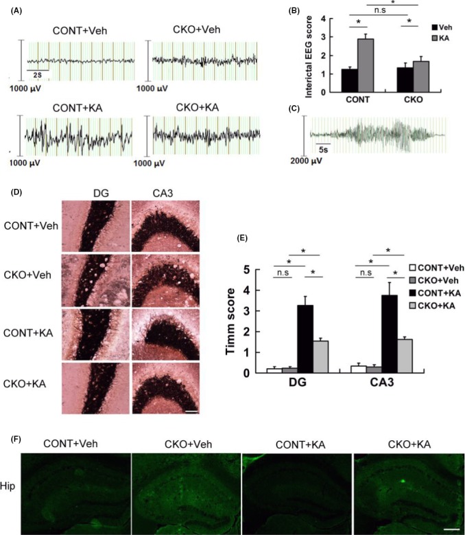
Epilepsy‐associated manifestation was attenuated in Rptor CKO mice. Control and Rptor CKO mice were treated with KA to induce status epilepticus and recorded with EEG. Mice were perfused and stained for mossy fiber sprouting 8 weeks after EEG recording. (A) Representative EEG background and interictal epileptiform spikes in different groups. (B) Score of EEG background spikes in different groups (n = 12 male mice per group). (C) Representative EEG depicting ictal activity from a spontaneous seizure. (D) Representative images of Timm staining in the dentate gyrus (DG) and CA3 zones (n = 6 male mice per group. Scale bar, 200 μm. (E) Quantitative summary of Timm staining. Robust mossy fiber sprouting was seen in KA‐treated control (CONT+KA) in the DG and CA3 zone, while it attenuated in Rptor CKO mice (CKO+KA) compared to their controls (CONT+KA or CKO+Veh). (F) Representative images of FJB staining 1 week after KA‐induced status epilepticus. No obvious cell death was noticed in all groups (n = 6 male mice per group). Scale bar, 10 μm. *P < 0.05 by one‐way ANOVA.
