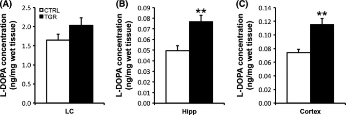
Figure 3.

In vivo TH activity is increased in the hippocampus and cortex of TGR rats. In vivo TH activity was estimated by measuring the regional accumulation of L‐DOPA in LC (A), hippocampal (B) and cortical (C) tissues extracted from control (white bars) and TGR rats (black bars) 20 min after the injection of the DOPA decarboxylase blocking agent NSD‐1015. Although TH activity tends to be increased in the LC region of TGR rats compared to controls, there is no significant difference between the two groups of rats. In contrast, TH activity is significantly increased in the hippocampus and frontal cortex of TGR rats compared to controls. Results are the mean concentration of L‐DOPA ± SEM (n = 5). **P < 0.01, ANOVA I.
