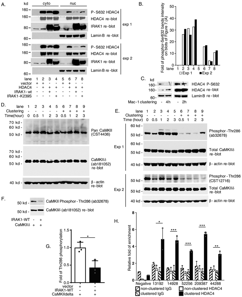
Figure 7. IL-1 receptor-associated kinase 1 (IRAK1) pathway via Calcium/calmodulin-dependent protein kinase II delta (CaMKIIδ) to downregulate histone deacetylase 4 (HDAC4) phosphorylation which facilitates HDAC4 recruitment to FOXP1 promoter during Mac-1 clustering.
(A) Immunoblot data of cytoplasmic and nuclear protein extracts from co-transfected HEK293 cells showing IRAK1-WT inhibits HDAC4 Ser632 phosphorylation. Two experiments were shown as exp 1 and exp 2. (B) Graph showing quantification of phosphor S632 HDAC4 bands from the blots shown in A. by ImageJ normalized to whole HDAC4. (C) Downregulation of HDAC4 Ser632 phosphorylation in nuclear extracts from THP-1 cells after 2 or 4 hours Mac-1 clustering. (D) Protein expression of CaMKIIδ in Mac-1 clustered THP-1 cells. (E) Mac-1 clustering inhibits Thr286 phosphorylation of CaMKIIδ. (F) IRAK1-WT inhibits Thr286 phosphorylation of CaMKIIδ in co-transfected HEK293 cells. (G) Summarized results of separate immunoblot experiments represented by F as graph showing quantification of phosphor-Thr286 CaMKIIδ bands on blots images analyzed by ImageJ normalized to total CaMKIIδ controls. (H) Enhanced recruitment of HDAC4 to P1 chromatin 2 hours after Mac-1 clustering detected by ChIP. Enriched amplicons were detected by quantitative PCR with 44/ak, 45/al, 46/am, 47/an and 48/aq (Supplementary Table). Immunoblot results of 2 hours and 4 hours after Mac-1 clustering in C represent 2 and 4 separate Mac-1 clustering/nuclear protein extraction/immunoblot experiments respectively. Results are n = 3 separate co-transfection/immunoblots experiments for G. Data represent the mean ± SD. *, P<0.05; **, P < 0.01; ***, P<0.001. Two tailed T-test.

