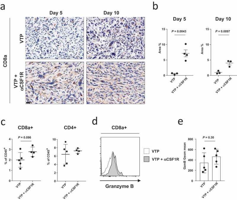
Figure 4.

Anti-CSF1R treatment increases CD8+ T cell infiltration in VTP-treated Myc-Cap tumors.
(a) Representative tumor section images of CD8+ T cell staining from tumors 5 and 10 days after VTP therapy alone or VTP therapy with anti-CSF1R. Scale bars represent 20 µm and (b) quantification of intratumoral CD8+ T cell from (a) as a percentage of area covered on days 5 and 10 after treatment. (c) Quantification of CD8+ and CD4+ T cells as a percentage of live CD45+ cell 10 days after treatment as assessed by flow cytometry. (d) Expression of granzyme B in CD8+CD45+ T cells as assessed by flow cytometry on day 10 after treatment and (e) quantification of granzyme B expression in CD8+CD45+ T cells from d). Data shown are a single representative of experiments performed at least twice.
