Figure 1.

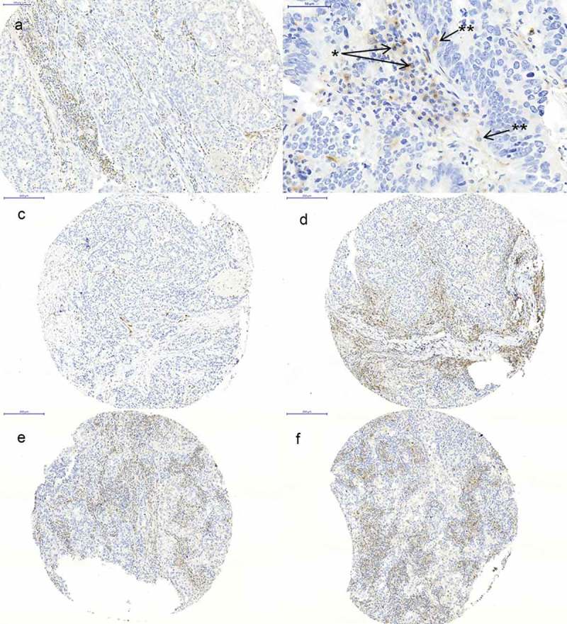
Immunohistochemistry of VISTA. (a) high expression of VISTA on TILs; (b) VISTA expression on lymphocytes (*) and macrophages (**); (c+d) tumor spots of the same tumor showing heterogeneous low and high VISTA expression; (e+f) tumor spots of the same tumor showing homogeneous VISTA expression.
