
Figure 1.
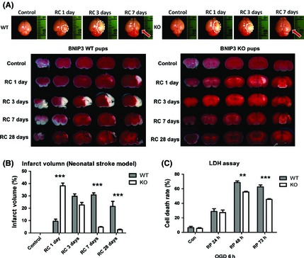
BNIP3 gene silencing was neuroprotective in stroke models. Measurement of brain infarct volume in BNIP3 WT and KO mice pups in neonatal stroke model. Two‐way ANOVA analysis and Bonferroni posttests were used to compare the total brain infarct volumes between the WT and KO groups: WT versus KO on each time point, ***P < 0.001. Control groups were sham‐operated and were not subjected to I/H treatment. N = 3–6 for each group (A, B). Time course of cell death rate in OGD/RP‐challenged BNIP3 WT and KO neurons, quantified by LDH assay. Two‐way ANOVA analysis and Bonferroni posttests were used to compare the cell death rates between WT and KO groups: WT versus KO on each time point, **P < 0.01, and ***P < 0.001. Control group were without I/H. N = 3 for each group (C).