
Figure 5.
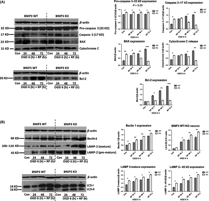
BNIP3 gene silencing decreased apoptosis and increased general autophagy in cortical neurons after OGD/RP injury. Decreased apoptosis was quantified by the decreased expression of pro‐apoptotic proteins (caspase 3, BAX and cytochrome c) and anti‐apoptotic protein (Bcl‐2) in the BNIP3 KO neuron (A). Increased general autophagy was quantified by the increased expression of autophagy marker proteins (Beclin1, LAMP‐2, and LC3‐II/I ratio) in the BNIP3 KO neuron (B). β‐Actin (43 kDa) was included as internal control. Band densities were measured by Quantity One software. Two‐way ANOVA analysis and Bonferroni posttests were used to compare the WT and KO groups: WT versus KO on each time point, *P < 0.05, **P < 0.01, and ***P < 0.001. N = 3 for each group.