
Figure 6.
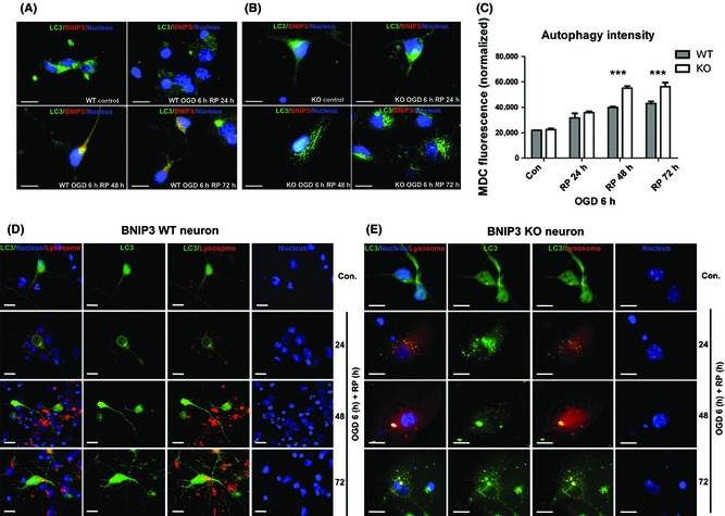
BNIP3 gene silencing increased general autophagy in OGD/RP‐challenged neurons. Immunocytochemistry was used to demonstrate the expression of BNIP3 and processing and translocation (punctuate staining) of LC3 protein. BNIP3 was stained with red, and LC3 and nuclei were marked with green and blue, respectively. Scale bars = 30 μm. Images were taken at 63× objective (A, B). MDC fluorescence was measured to quantify the general autophagy intensities in BNIP3 WT and KO neurons. Two‐way ANOVA analysis and Bonferroni posttests were used to compare the WT and KO groups: WT versus KO on each time point, ***P < 0.001. N = 3 for each group (C). Co‐localization of lysosomes with autophagosomes inside BNIP3 WT and KO neurons was detected. Lysosomes were stained with red, and LC3 and nuclei were marked with green and blue, respectively. Scale bars = 30 μm. Images were taken at 63× objective (D, E). Control groups without OGD/RP.