
Figure 1.
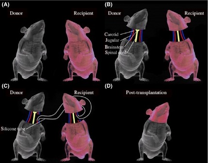
Schematic representation of the AHBR mouse model. (A) Two mice with different colors are selected. (B) All tissues have been separated including carotid artery, jugular vein, spinal cord and so on. (C) In order to ensure transplanted brain tissue does not stop blood circulation to avoid cerebral ischemia and hypoxia, cross‐circulation is established by the silicone tubes. (D) Mice after transplantation.