Figure 4.

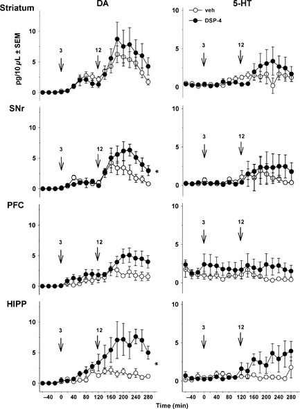
Effect of DSP‐4 on the extracellular levels of dopamine (DA) and 5‐HT induced by l‐DOPA in 6‐OHDA‐lesioned rats. Data represent mean + SEM (n = 5–6 rats/group) of extracellular concentration (pg/10 μL dialysate) of DA (left panel) and 5‐HT (right panel) versus time, observed in striatum, substantia nigra pars reticulata (SNr), prefrontal cortex (PFC) and hippocampus (HIPP). Microdialysis experiments were performed 3–4 weeks after the unilateral injection of 6‐OHDA. One week before the microdialysis experiments, 6‐OHDA‐lesioned rats received an intraperitoneal (i.p.) administration of 50 mg/kg DSP‐4 or its vehicle (veh). l‐DOPA was administered i.p. at 3 mg/kg (time 0, first arrow) and was preceded 20 min before by the administration of benserazide (15 mg/kg, i.p.). Two hours after the first l‐DOPA injection, rats received another i.p. injection of l‐DOPA at 12 mg/kg (time 120, second arrow). Asterisks refer to the probability level of statistical significance of the overall effect of l‐DOPA at 12 mg/kg in the DSP‐4 group versus the veh group: *P < 0.05 (Student's t‐test).
