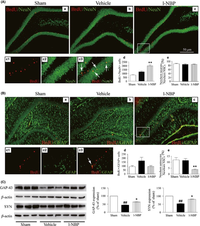
Figure 4.

L‐NBP increased the number of newborn neurons and the expressions of GAP‐43 and SYN but decreased the percentage of newborn GFAP‐positive cells. Representative confocal fluorescence images immunolabeled with BrdU (red) and NeuN (green) or GFAP (green) antibodies, BrdU+/NeuN+ cells shown yellow color (arrow) (A), and BrdU+/GFAP + cells shown yellow color (arrow) (B). Quantitative analysis of BrdU+/NeuN+ (A‐d) and BrdU+/GFAP + cells (B‐d), the percentage of BrdU+/NeuN+ (A‐e) and BrdU+/GFAP + (B‐e). Western blots of GAP‐43 and SYN in the hippocampus of injured hemisphere treated with vehicle or l‐NBP, respectively (C). Quantified results were normalized to β‐actin expression. Values were expressed as percentages compared to sham‐operated rats (set to 100%) and represented as group mean ± SEM. n = 12–13 rats per group. ## P < 0.01 versus sham group, *P < 0.05, **P < 0.01 versus vehicle‐treated group.
