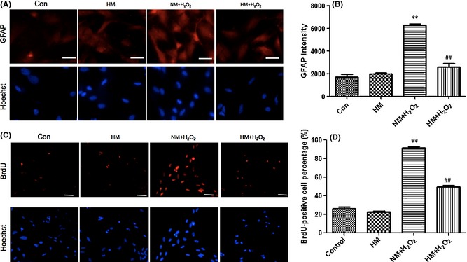
Figure 5.

Twelve hours after adding H2O2 (100 μM) and FeCl2 (15 μM) to the medium, with or without super‐saturated hydrogen, the hypertrophy and proliferation of cultured astrocytes were observed. (A) and (B) Immunocytochemistry of glial fibrillary acidic protein (GFAP) demonstrating the morphological characteristics of hypertrophy and excessive expression of GFAP, and the fluorescence intensity data. (C) and (D) Images demonstrating the DNA synthesis of BrdU, which reflects the proliferation of astrocytes, and the percentage of positive cells. Hypertrophy, proliferation, and excessive expression of GFAP were obviously induced by H2O2 in the normal medium (NM) + H2O2 group (vs. control, P < 0.01) and were significantly inhibited by treatment with hydrogen‐rich medium (vs. NM + H2O2, P < 0.01). Results were means ± SEM of three independent experiments. **P < 0.01, NM + H2O2 vs. control; ##P < 0.01, H2O2 + hydrogen‐rich saline vs. H2O2 + normal saline. Scale bar for A = 20 μm and for C = 50 μm.
