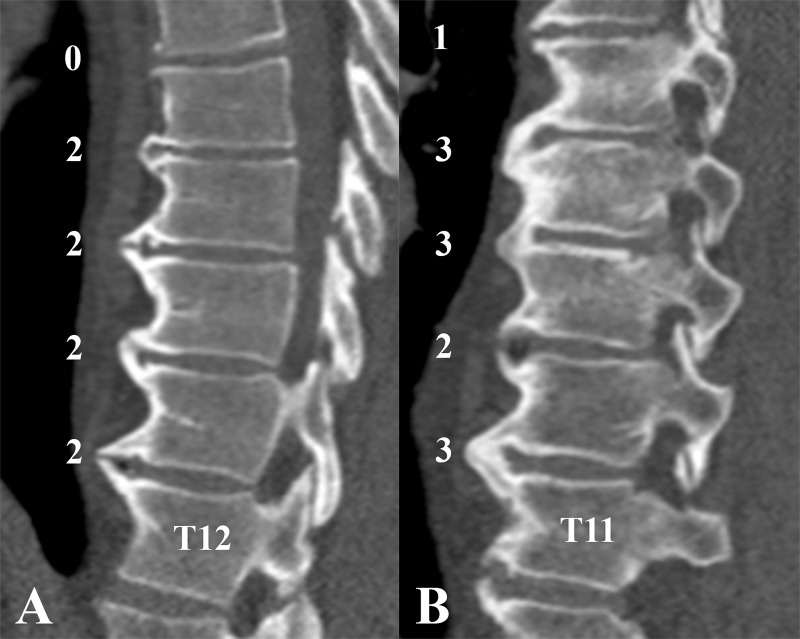
Figure 4:
Example images in patients with early diffuse idiopathic skeletal hyperostosis (DISH). A, Sagittal CT image in a 54-year-old man with early DISH with grades based on the flowchart shown in Figure 3. No complete bridge was present at any spinal segment; however, four segments were scored as showing a near-complete bone bridge (three or more segments with a score of 2). B, Sagittal CT image in a 67-year-old man with early DISH is shown. Three complete bone bridges (score of 3) were present, but not in an adjacent fashion, as is required to fulfill the criteria for DISH of Resnick and Niwayama (7). According to the flowchart in Figure 3, a complete bone bridge was present, and the highest bridge score of an adjacent segment was also a complete bone bridge (score of 3). Adjacent to these two complete bridge scores, the highest bridge score was a near-complete bone bridge (score of 2), and thus no adjacent three segments with complete bridges were present, excluding definite DISH and resulting in the diagnosis of early DISH. See also Movie 1 (online).

