
Figure 1.
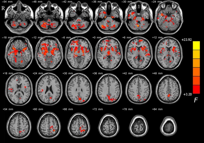
Brain maps for amplitude of low‐frequency fluctuations (ALFF) differences among tremor‐dominant (TD), postural instability/gait difficulty (PIGD), and healthy control (HC) groups. The statistical significant level was set at P < 0.05 and cluster size >237 voxels within the group mean gray mask, which corresponded to a corrected P < 0.05. The left side of the image corresponds to the right side of the brain in axial orientation; slice coordinates according to Montreal Neurological Institute (MNI) space are shown in the upper right corner of the slices, indicating Z‐axis in axial orientation.