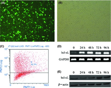
Figure 1.

Overexpression of Bcl‐xL in SH‐SY5Y cells. (A) The transfected cells were analyzed under a fluorescent microscope. (B) The morphology of SH‐SY5Y cells was observed under bright field microscope. (C) Transfection efficiency was detected 48h after transfection by flow cytometry. (D) RT‐PCR and (E) Western blotting were employed to detect the mRNA and protein expression of Bcl‐xL 24, 48, 72, and 96h after transfection, respectively.
