
Figure 3.
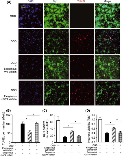
Effects of exogenous recombinant wild‐type (WT) visfatin (300 ng/mL) and H247A‐mutant visfatin (300 ng/mL) on neuron in OGD model. (A) Representative images of mediums of neuron in OGD model (2 h). Tuj‐1 (green) was a marker of neuron axon. TUNEL (red) was used to detect apoptosis. DAPI was used to stain nucleus. (B) Quantitative analysis of TUNEL‐positive apoptotic neurons. *P < 0.05, n = 6. (C) Quantitative analysis of Tuj‐1‐positive survival neurons. *P < 0.05, n = 6. (D) Cell viability assay on neurons. *P < 0.05, n = 6.