
Figure 3.
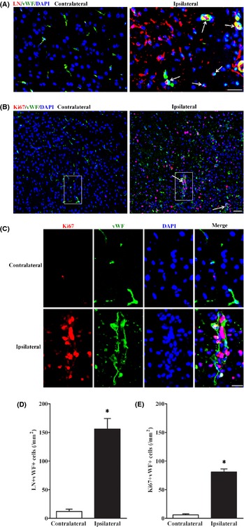
Some of the laminin‐positive cells were endothelial cells in the IBZ after pMCAO. (A) LN/vWF double staining showed part of laminin (LN)‐positive cells in IBZ at day 7 after pMCAO were endothelial cells (arrows). Scale bar = 20 μm. (B) Combination staining of antibodies against an endothelial cell marker (vWF) with a proliferation marker (Ki67) showed that endothelial cells formed new vessels in IBZ at day 7 after pMCAO. Scale bar = 20 μm. (C) is magnified images of boxes in (B). Scale bar = 10 μm. (D) Quantification of LN/vWF dual positive cells in IBZ 7 days after pMCAO. (E) Quantification of Ki67/vWF dual positive cells in IBZ 7 days after pMCAO. n = 7–8/group. *P < 0.05, versus. contralateral side.