
Figure 4.
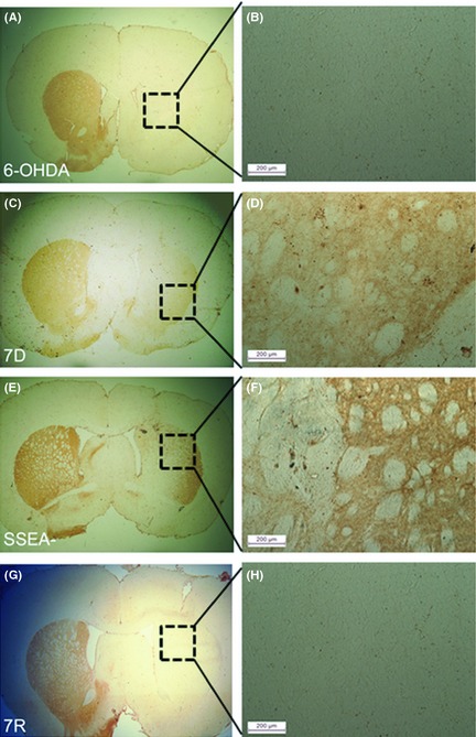
Tyrosine hydroxylase (TH) immunoreactivity in the striatum. (A) Ipsilateral side of the 6‐OHDA control group showed no TH staining in the dorsolateral striatum, and (B) shows the magnified image of the dotted square in the image, A. Striatal sections from rat brains transplanted with cells from mES differentiated for 7 days (7D) (C); 7D cells free of the dividing cells (SSEA−) (E). mES differentiated with serum‐free media and retinoic acid (7R) (G) did not show the presence of TH neurons in the graft. Magnification 4×. Magnified images of the area within the dotted squares of A, C, E and G, that is, B, D, F and H, respectively, showed the presence TH‐stained fibers in the ipsilateral striatum of 7D (D) and SSEA− (F) cell‐transplanted groups (Scale bars: 200 μm).