
Figure 2.
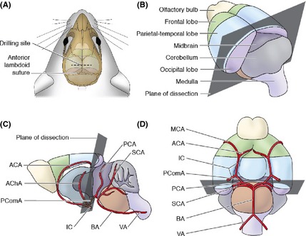
Location of the cutting plane in donor mice. (A) Drill 3 mm distal to the anterior lambdoid suture, (B) superolateral aspect of the brain showing the cutting plane, (C) sagittal section, and (D) inferior aspect of the brain showing the cutting plane and vasculature. The cutting plane passes from the caudal edge of the parietal–temporal lobes, along the rostral aspect of the midbrain, and through the PcomA. This preserves blood flow to the regions of the donor's brain that are retained. ACA, anterior cerebral artery; AChA, anterior choroidal artery; PcomA, posterior communicating artery; IC, internal carotid; MCA, middle cerebral artery; PCA, posterior cerebral artery; SCA, superior cerebellar artery; BA, basilar artery; and VA, vertebral artery.