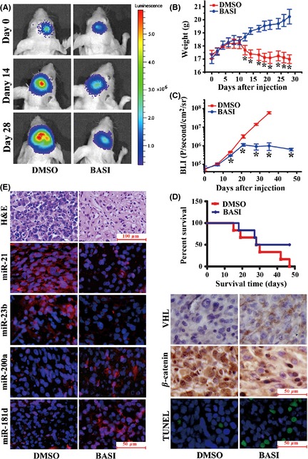
Figure 6.

Antitumor effects of BASI in a U87 orthotopic intracranial model. (A) Representative pseudocolor bioluminescence images of mice that were implanted with intracranial tumors and treated intraperitoneally with 40 mg/kg BASI or DMSO on days 0, 14, and 28. (B) A plot depicting the Fluc activity measured bioluminescence imaging for the BASI and DMSO treatment groups. Data represent the mean ± SD (*P < 0.05). (C) A plot showing the body weight changes of nude mice bearing U87 orthotopic tumors. Weights were measured every 2 days following the intraperitoneal injection. Data represent the mean ± SD (*P < 0.05). (D) The overall survival of mice in the DMSO and BASI treatment groups for this same experiment. There was a substantial survival benefit for the BASI‐treated mice. (E) Representative photomicrographs of H&E staining, FISH for miR‐21, miR‐23b, miR‐200a, and miR‐181d, immunohistochemistry (IHC) for VHL and β‐catenin, and TUNEL staining on orthotopic tumor sections.
