
Figure 4.
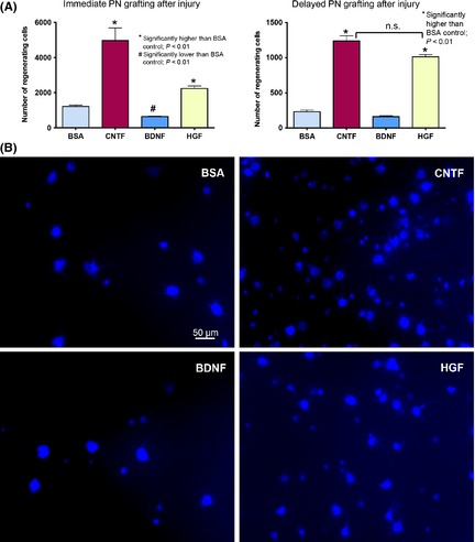
(A) Graphs comparing the number of ganglion cells that have regenerated axons into a PN graft under the influence of trophic factors. Left panel shows the result from immediate PN grafting after optic nerve cut and injection of growth factors, and right panel illustrates the results obtained when PN grafting was delayed for 7 days after optic nerve cut. n.s. not significantly different between the CNTF and HGF groups when PN grafting was delayed. (B) Photomicrographs depicting ganglion cells in the wholemount retina that have regenerated axons into a PN grafted to the cut optic nerve immediately after injury and injection of trophic factors. The regenerating cells have been retrogradely labeled by the fluorescent dye GB applied to the PN graft at 25 days after grafting and the retina examined 3 days later (at 28 days post‐grafting). Note that ganglion cells of different sizes have regenerated into the graft in each group, and that HGF and CNTF stimulated more ganglion cells to regenerate (~ 2 and 4 times, respectively, of BSA control), while BDNF reduced regeneration.