
Figure 7.
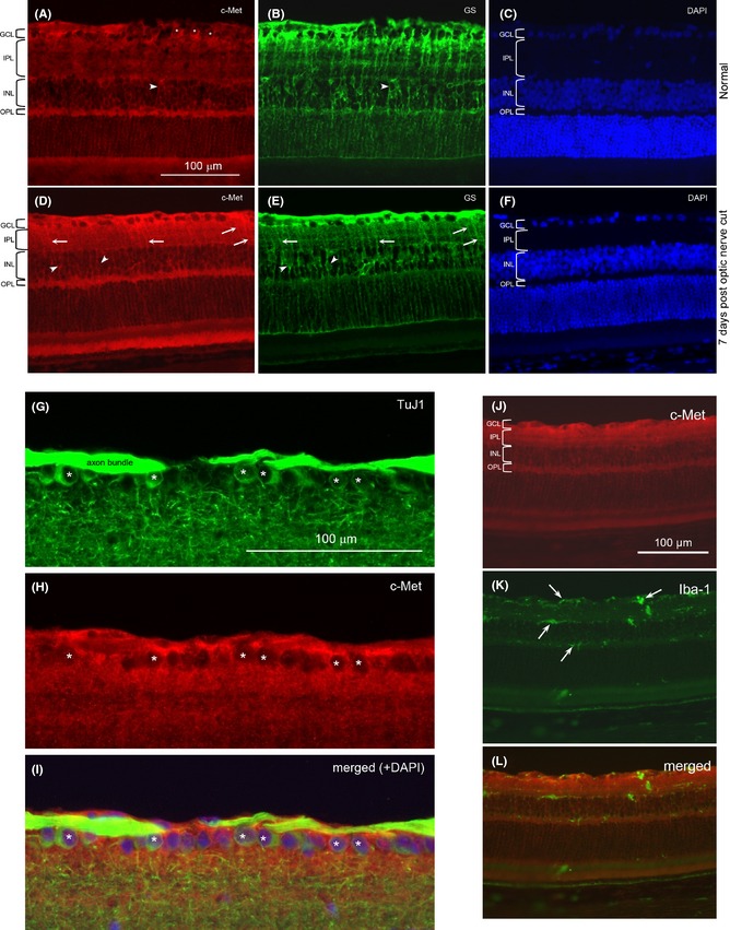
C‐Met localization in the retina. A.‐F. Double staining of retinal sections with anti‐c‐Met (red) and anti‐glutamine synthetase (GS, green), and counterstained with DAPI nuclear stain: from a normal retina (A‐C) or retina at 7 days post‐optic nerve cut (D‐F). The various retinal layers have been marked: GCL: ganglion cell layer; IPL: inner plexiform layer; INL: inner nuclear layer; OPL: outer plexiform layer. Asterisks (*) indicate c‐Met‐labeled cell bodies in the GCL that are confirmed to be ganglion cells by double‐labeling with TuJ1 (see G‐I below). Arrowheads point to cell bodies in the INL that belong to Muller cells, while the arrows point to stained processes in the IPL that belong to radial processes of Muller cells. Note the increase in anti‐c‐Met staining after optic nerve injury, especially in GCL and IPL. The c‐Met‐labeled radial processes of Muller cells in the IPL are also more prominent after optic nerve cut. G.‐I. Double staining of TuJ1 and anti‐c‐Met from a normal retina. The micrographs showed the GCL and part of the IPL to illustrate that many ganglion cells labeled by TuJ1 (marked by *) in G are also positive for anti‐c‐Met in H. The merged view of the 2 labeling together with DAPI counterstain is shown in I. The apparent “emptiness” of the c‐Met‐labeled cells in H is due to the fact that c‐Met is localized on the cell membrane so that the red label surrounds the unstained cytoplasm. J.‐L. Double staining of a retinal section at 7 days post‐optic nerve cut with anti‐c‐Met and anti‐Iba‐1. Note that the Iba‐1‐stained cells (arrows) do not have any correspondence with the anti‐ c‐Met staining, showing that microglia do not express c‐Met.