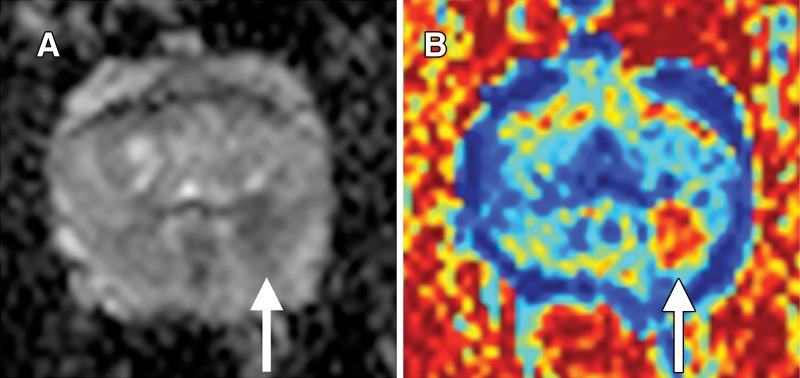
Figure 4:
Images in a 57-year-old man with targeted biopsy–proven Gleason 3+4 prostate cancer. A, Apparent diffusion coefficient (ADC) map shows reduced ADC in left peripheral zone at 3 to 5 o’clock (arrow). B, Vascular, Extracellular, and Restricted Diffusion for Cytometry in Tumors (VERDICT) intracellular volume fraction map. Tumor (arrow) is very conspicuous.

