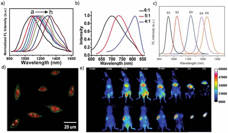
Figure 4.

(a) PL spectra of Ag2Se QDs with different reaction times (b) PL spectra of monodispersed Ag2Se QDs with different sizes (molar ratio of Ag: Se = 6:1, 5:1, and 4:1). (c) Emission spectra of Ag2Se QDs capped with a multidentate polymer (samples S1−S5 corresponding to 0.5, 1, 1.5, 2, and 3 h reaction). (d) Overlay image (confocal laser scanning microscopy and NIR) of the Cu-2-{2-chloro-6-hydroxy-5-[(2-methyl-quinolin-8-ylamino)-methyl]-3-oxo-3H-xanthen-9-yl}-benzoic acid (CuFl) stained cells after incubation with the Ag2S-GSH-SNO NPs for 3 h. (e) In vivo imaging of PEGylated Ag2Se QDs in mice after intravenous injection. (Top row) Abdomen imaging; (bottom row) backside imaging. Reproduced with permission from (Figure 4(a), [52] Copyright 2013 The American Chemical Society; Figure 4(b), [60] Copyright 2012 The American Chemical Society; Figure 4(c), [54] Copyright 2014 The American Chemical Society; Figure 4(d), [53] Copyright 2013 The American Chemical Society; and Figure 4(e), [ 48] Copyright 2016 The American Chemical Society).
