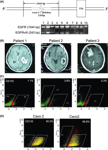
Figure 1.

RT‐PCR results showed that all 10 GBM clinical specimens were wild‐type EGFR +, and eight of the 10 were EGFRvIII + (A). Lanes correspond to the patient number. DNA fragments of 1044 bp and 243 bp in size represented EGFR and EGFRvIII, respectively. MRI showed space‐occupying lesions in all three patients (B). Patient No. 1. Space‐occupying lesions were located in the left temporal lobe rear, and the midline structure shifted right. Patient No. 2 showed a class round abnormal signal shadow, and rich blood supply space‐occupying lesions were located in the left temporal lobe rear. Patient No. 3 showed space‐occupying lesions in the right frontal lobe, the right lateral ventricle was compressed by the tumor, and the midline structure shifted left. Flow cytometric analysis showed that the percentages of CD133+ cells from each patient were 1.1%, 3.6%, and 0.3% (C). The Caco2 cell line with CD133 overexpression was used as a positive control (D).
