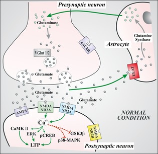
Figure 1.

This figure represents the physiological glutamate‐mediated transmission at synaptic level. Glutamate release from presynaptic terminal acts through the activation of ionotropic glutamate receptors located in postsynaptic terminal. In the figure is in particular described the N‐methyl‐d‐aspartate (NMDA) signaling. The activated NMDA NR2A induces increase in calcium, which in turn favors the activation of several metabolic pathways (CaMK, ERK, and CREB) responsible for anabolic activation with subsequent activation of long‐term potentiation (LTP) mechanisms. Conversely, the calcium increase inhibits metabolic pathways (GSK3β and MAPK) responsible for long‐term depression and synaptic remodeling. Glutamate excess is transported via the EAAT into astrocytes where it is transformed to glutamine via the synthase activity which then again returns to synaptic terminals where the enzyme glutaminase produces again glutamate. The produced glutamate is subsequently filled in vesicles through a specific transporter (VGlut). VGlut (vesicular glutamate transporter); EAAT (excitatory amino acid transporter); α7‐nAchR (alpha‐7 nicotinic acetylcholine receptor); NMDANR2A (N‐methyl‐d‐aspartate NR2A subunit); NMDANR2B (N‐methyl‐d‐aspartate NR2B subunit); ERK (extracellular signal‐related kinase); CaMKII (calcium calmodulin‐dependent kinase II); pCREB (phosphorylated cyclic AMP response element binding protein); GSK3β (glycogen synthase kinase 3β); p38‐MAPK (p38 mitogen‐activated protein kinase).
