
Figure 3.
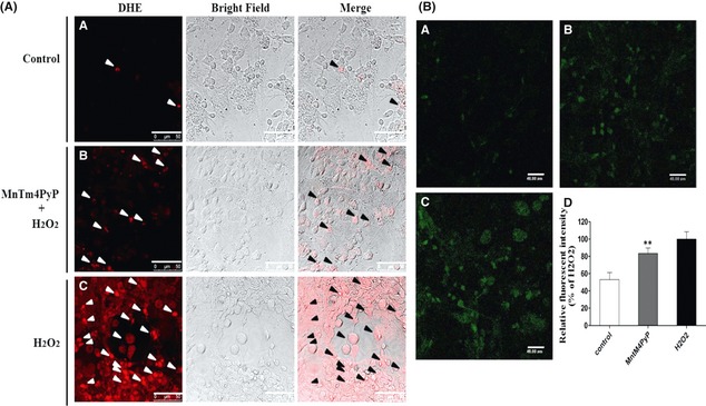
MnSOD mimic eliminates intracellular superoxide radical and H2O2 levels. (A) Representative images of laser scanning confocal microscopy. Arrows indicate red fluorescence representing the presence of intracellular superoxide radical. (B) Representative images of laser scanning confocal microscopy. Green fluorescence represents intracellular H2O2 levels. (B‐A) Cells treated with PBS; (B‐B) cells treated with 5 μM MnTm4PyP before H2O2 (100 μM) exposure for 2 h; (B‐C) cells treated with 100 μM H2O2 for 2 h; (B‐D) 0 Quantification of fluorescence intensity from mean ± SD, (n = 6). **P < 0.01 compared with the H2O2 group.