
Figure 4.
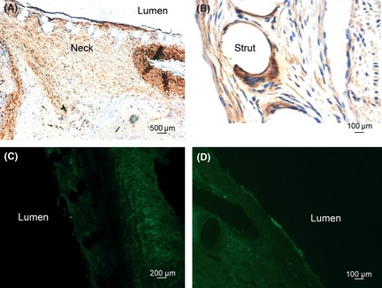
Histological analysis of neointima at 2 weeks and 4 weeks after FD treatment. At 2 weeks after FD treatment, immunochemistry shows that cells with positive α‐actin staining are distributed in the neointima of the aneurysm neck (A); cells with positive CD68 staining are located around the stent struts (B). Few cells with positive vWF staining are observed on the surface of neointima (C). At 4 weeks after FD treatment, cells with positive vWF staining are observed on the surface of neointima (D).