
Figure 1.
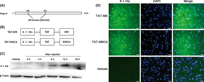
Construction and transduction of trans‐activator (TAT)‐M9 fusion proteins. (A) The structure of the M9 domain. (B) The construction of TAT‐M9CA and TAT‐M9. (C) Western blot showing TAT‐M9 uptake in the brain after intraperitoneal injection. (D) TAT‐M9 was double‐stained with the nuclear marker DAPI and rapidly taken up into the brain. TAT‐M9 showed widespread uptake in the brain using anti‐6 × His antibody to test the signal 6 h after intraperitoneal injection compared with vehicle. TAT‐M9CA showed similar results. Scale Bar = 20 μm.